Overview of Themes for the Patient Community
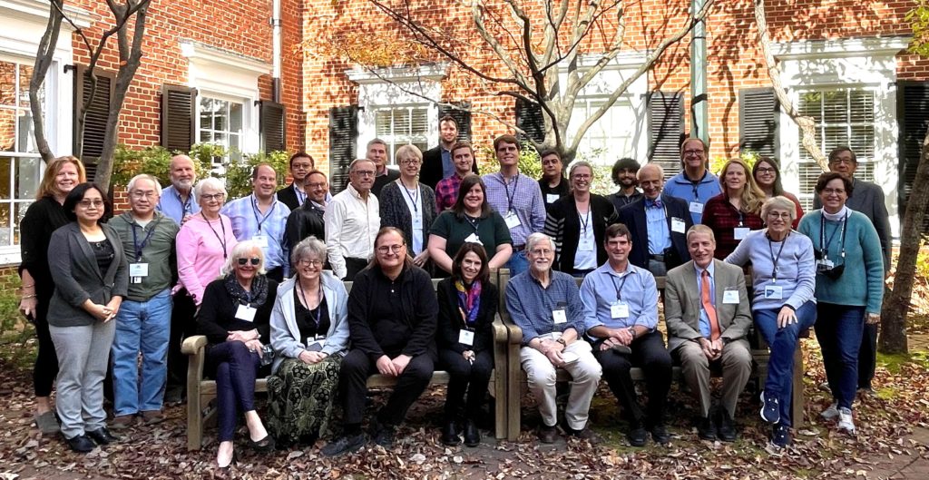
The 2021 ANA Symposium: PAX6, Aniridia, and Beyond was held November 4-6 on the campus of the University of Virginia. The meeting was attended by scientists, clinicians, and representatives of patient advocacy organizations, and addressed aniridia-related topics, including corneal health, genetics, eye development, glaucoma, and research models and techniques.
This Symposium was a forum for scientists and medical professionals to discuss their work with each other. The purpose of this overview is to share patient-relevant information from these discussions.
A printable version of this post is available in English in regular print and in enlarged print. To read this post in a language other than English, please use the translate function at the top of this page.
Hope for the Future: Corneal Treatment
Following are overviews of presentations focused on the aniridic cornea:
Daniel Aberdam (Université de Paris, France) is exploring drug-repurposing using a stem cell model to test thousands of drugs already on the market. This process identified two compounds that increased PAX6 expression. If this effect is confirmed in the ongoing animal testing, then these drugs could potentially be tested quickly in clinical trials in humans.
Ali Djalilian (University of Illinois, USA) also showed his latest data using a drug-repurposing approach to enhance PAX6 expression. He also provided an update on early stage clinical trials using Mesenchymal Stromal Cells for promoting corneal repair.
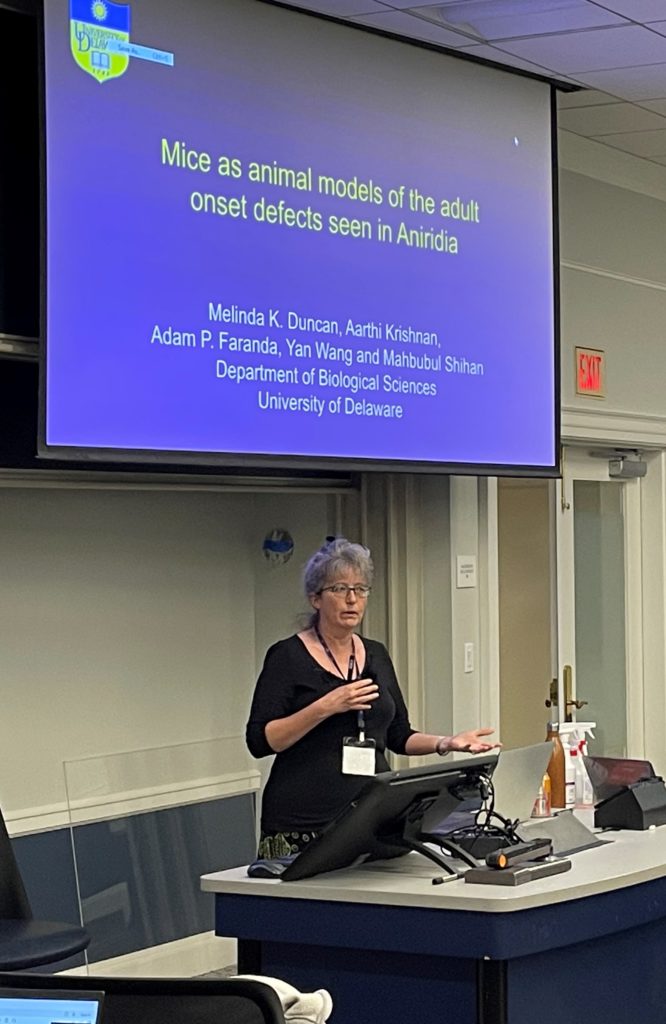
Melinda Duncan (University of Delaware, USA) described a study of the progressive loss of corneal transparency in mice, which helped to better understand defects in the aniridic cornea. This study revealed several potential therapeutic targets for corneal disease in aniridia that will require further investigation.
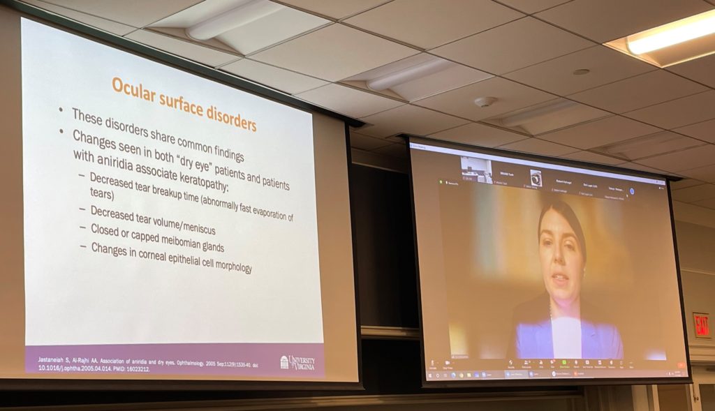
Tara McGehee (University of Virginia, USA) described a man-made version of lacritin, which is a protein found naturally on the surface of the eye. This synthetic protein is being tested in people with another type of ocular surface disease. If it works well, it may be useful for treating the cornea in aniridia.
Elizabeth Simpson (University of British Columbia, Canada) discussed the potential that modifying or “editing” genes using the technique CRISPR-Cas9 holds for people with aniridia. She emphasized that you have to cure the mouse before you can cure the human. With gene therapy, the mouse hasn’t been cured yet, but once it is, it takes an average of four years to bring the therapy to clinical trials.
Multiple presentations focused on the desire to find better treatments for aniridia-associated keratopathy (AAK). AAK is a progressive condition in a majority of people with aniridia that causes pain and vision loss. Few medical treatment options exist for management of AAK, and surgical options are invasive and carry high risks of complications.
It was encouraging to learn that multiple different avenues are being explored to address corneal problems experienced by people with aniridia. Many of these potential treatments could be used regardless of the patient’s genetic mutation or deletion.
New Information Impacting Patient Care

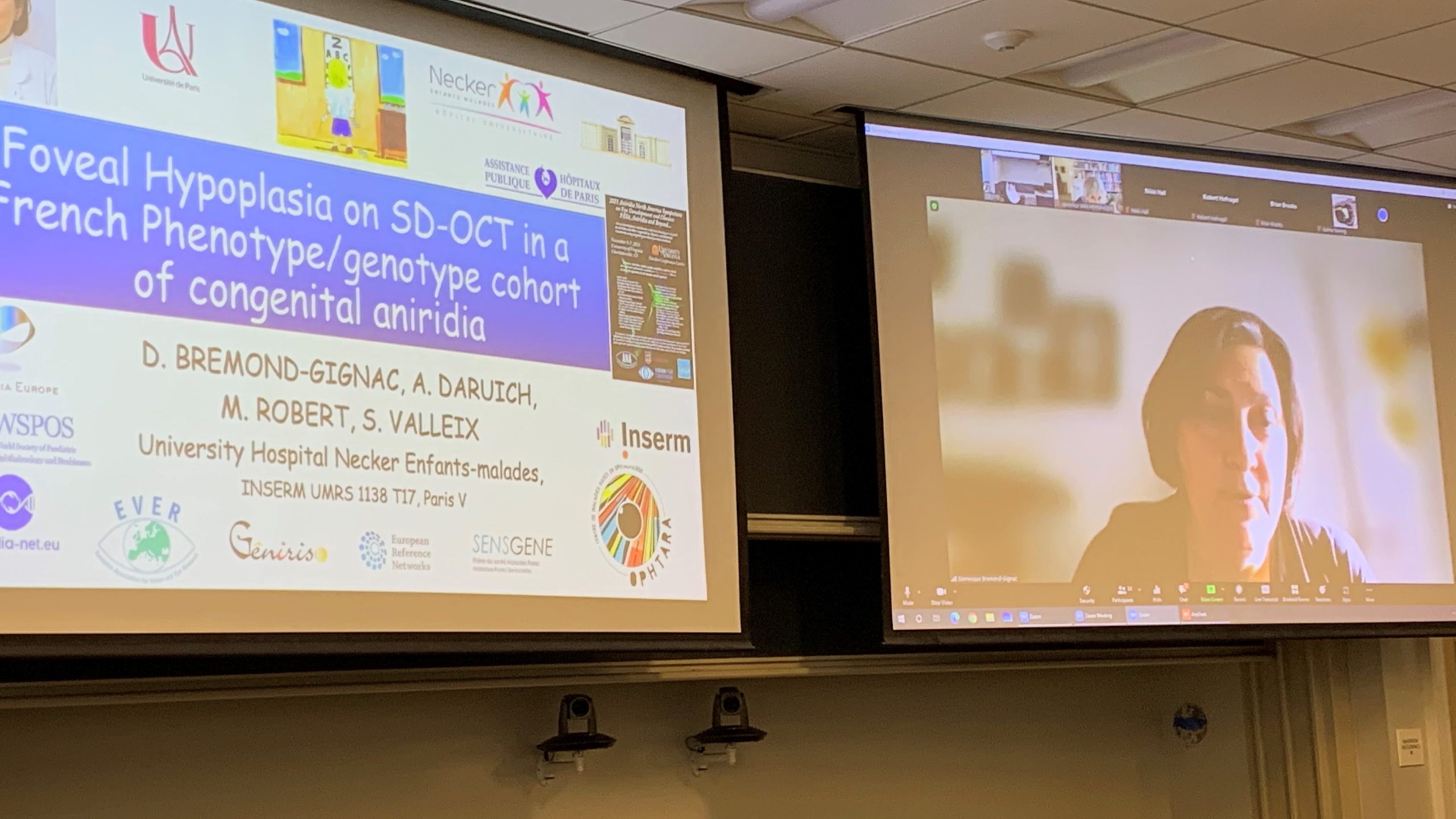
Dominique Bremond-Gignac (Université de Paris, France) presented a study comparing foveal and iris hypoplasia (phenotype) to specific genetic mutations (genotype). This information could have predictive value regarding visual acuity, and could also help determine whether future treatments or interventions could improve baseline visual acuity.
Mariya Moosajee (UCL Institute of Ophthalmology, UK) presented a study about the various mutation types that may affect the PAX6 gene and lead to aniridia. Some of these mutations can have a more severe effect on the eyes, while others have a milder effect. It’s important to classify these findings so when clinicians see new patients, they can advise regarding possible complications. In addition, the study looked at more than 1000 metabolites (such as proteins, carbohydrates, fats, and vitamins) from the blood of aniridia patients and identified changes that may influence weight, sleep, behavior, and mood.
Numerous presentations discussed research results that could directly impact patient care. These results included new studies linking the type of genetic mutation to the way aniridia presents in people with that type of mutation. It also included discussions of new surgical and imaging techniques, and proper timing of treatment to preserve corneal function. Comparison of differences in laboratory results and sleep disturbances in the aniridic versus non-aniridic populations was also covered.
It was reassuring to hear the practical information that came out of these studies and how this knowledge may alter patient care in the future.
Following are overviews of presentations impacting patient care:
Hao Zhang (Northwestern University, USA) presented a new type of imaging used to view abnormalities in the anatomy of the eye that can contribute to glaucoma. This new imaging, called vis-OCT, gives incredibly detailed images of the eye, making it a valuable tool in clinical settings.
Neil Lagali (Linköping University, Sweden) discussed the evidence for early treatment of the cornea in children with aniridia, because an attempt to support the normal corneal function could delay or halt the progression of AAK in adulthood. He also discussed the challenges involved in determining the appropriate dosing regimen, timing, and follow-up of potential treatments for clinical trials.
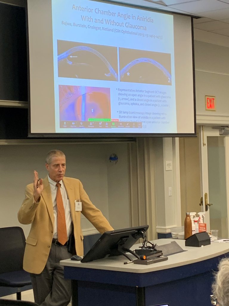
Peter Netland (University of Virginia, USA) explained the types of glaucoma and treatments for glaucoma in aniridia. There have been major improvements in the treatment of glaucoma, and their relevance to individuals with aniridia was discussed. New information about visual acuity in individuals with WAGR Spectrum was presented, demonstrating that vision is significantly worse in individuals with WAGR Spectrum when compared to those with isolated aniridia.
Amazing Intricacies of the Genetic Code
Many presenters touched on the topic of genetics in some way. This information emphasized how complex the gene networks are that govern eye development, as well as how even very small changes can make a very large difference. It was awe-inspiring to realize how much has been learned about the genetics of aniridia during the last 30 years.
Following are overviews of presentations related specifically to genetics:
Seth Blackshaw (Johns Hopkins University, USA) presented his work profiling single cell types over the full course of development in the human and mouse retina. This work identified many previously unknown genes that regulate retinal development and provide the basis for further precise understanding of retinal diseases arising in these cell types.
Ales Cvekl (Albert Einstein College of Medicine, USA) highlighted updates on his lab’s work with the PAX6 gene. His lab has developed methods that allow them to systematically probe the effects of individual human PAX6 mutations. In the future, these methods may be applied to create patient-specific treatments. This is an encouraging example of the possibilities of personalized medicine.
Nikki Hall (University of Edinburgh, UK) presented work doing whole genome sequencing on 37 aniridia patients whose genetic cause had previously been unknown. The talk discussed these cases as well as different ways to analyze the whole genome sequences.
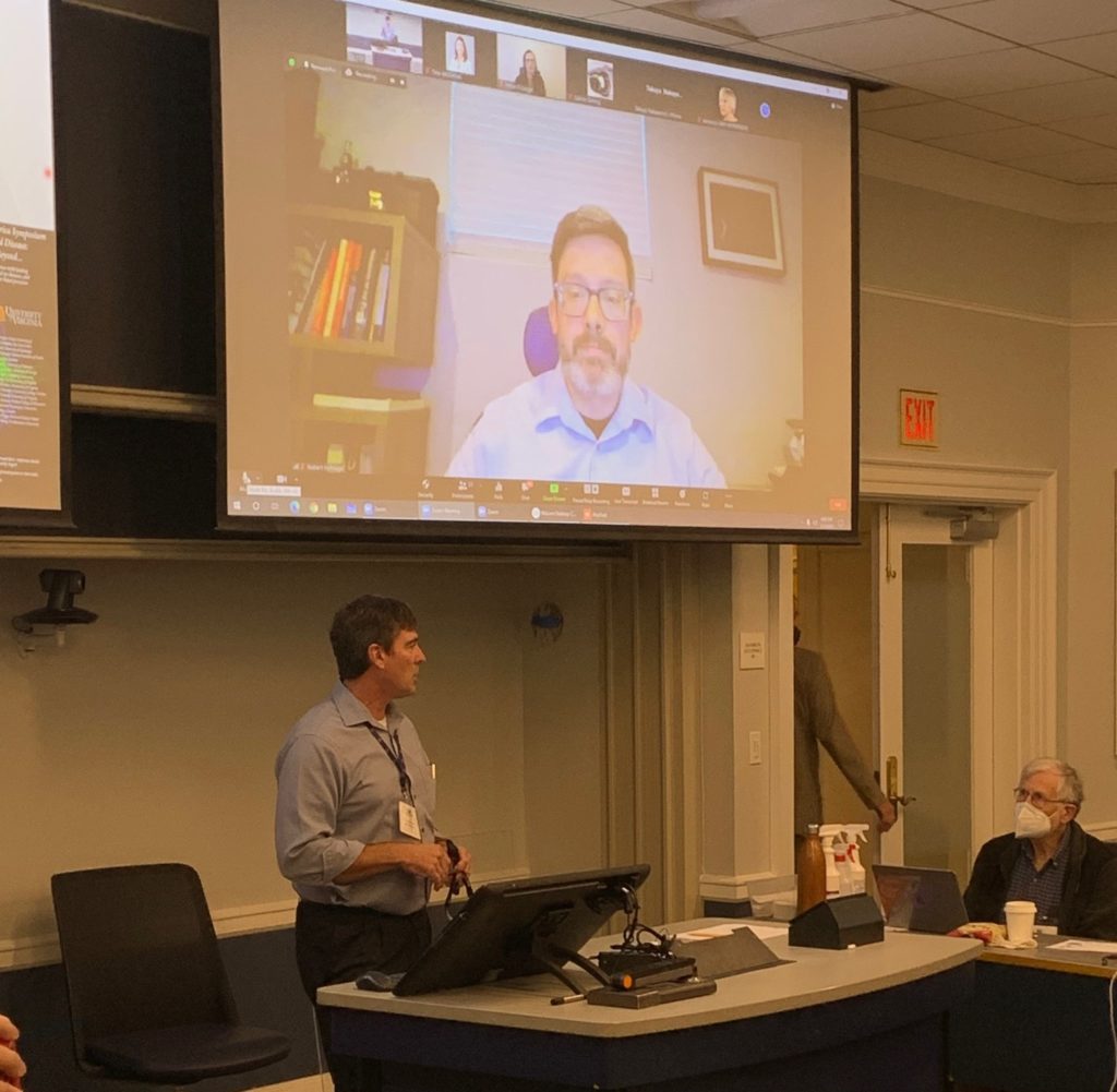
Robert Hufnagel (National Eye Institute, NIH, USA) presented an investigation of the genetic causes of foveal hypoplasia in patients seen at the National Eye Institute. Exome and genome sequencing was performed to identify the genetic basis in previously unsolved cases. This work contributes to our understanding of genetic pathways that contribute to foveal hypoplasia, a common feature of aniridia.
Elena Semina (Medical College of Wisconsin, USA) updated an ongoing study of unexplained cases of aniridia. Through this study, some new genes have been discovered to cause aniridia that were previously unknown.
Veronica van Heyningen (Institute of Genetics and Cancer, UK) talked about finding mutations in non-coding regions of the PAX6 gene and the importance of learning how these non-coding regions are involved in the expression of PAX6.
Janey Wiggs (Harvard Medical School, USA) discussed genetic research in glaucoma, which has led to the discovery of a number of genes and genetic risk factors that can contribute to disease.
Exciting New Techniques in Research
Various presentations addressed new techniques being explored in research on aniridia and its related conditions. Many of these techniques fall under the umbrella of “basic science” research, which is a crucial first step towards discovering new treatments.
Although this type of research is highly technical and often not directly applicable to patients, it is fascinating to see the new strategies being investigated to propel research forward.
Following are overviews of talks related to new techniques in research:
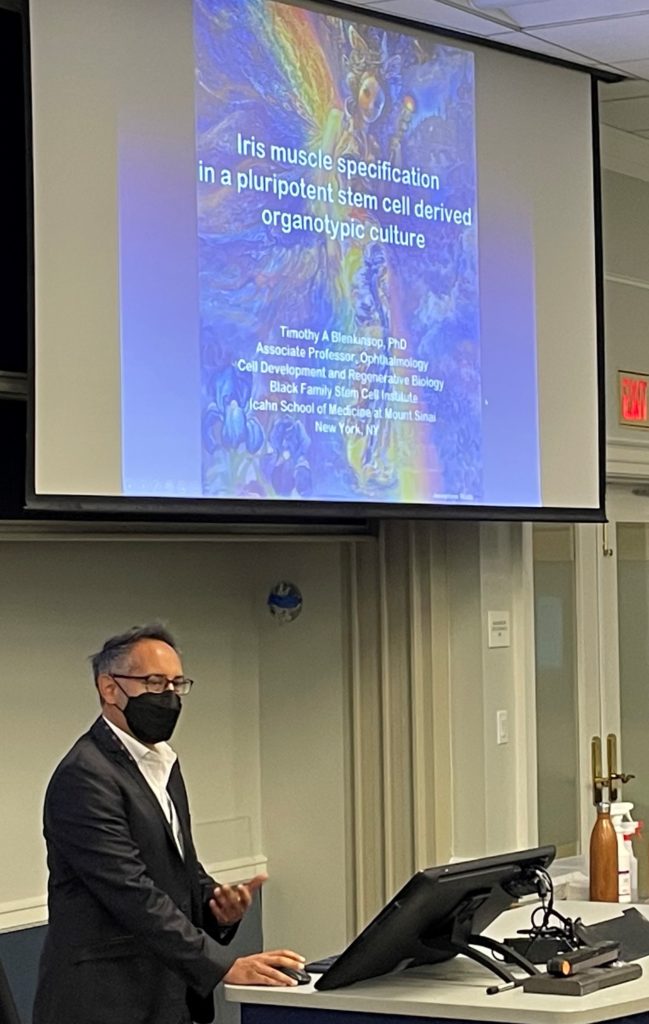
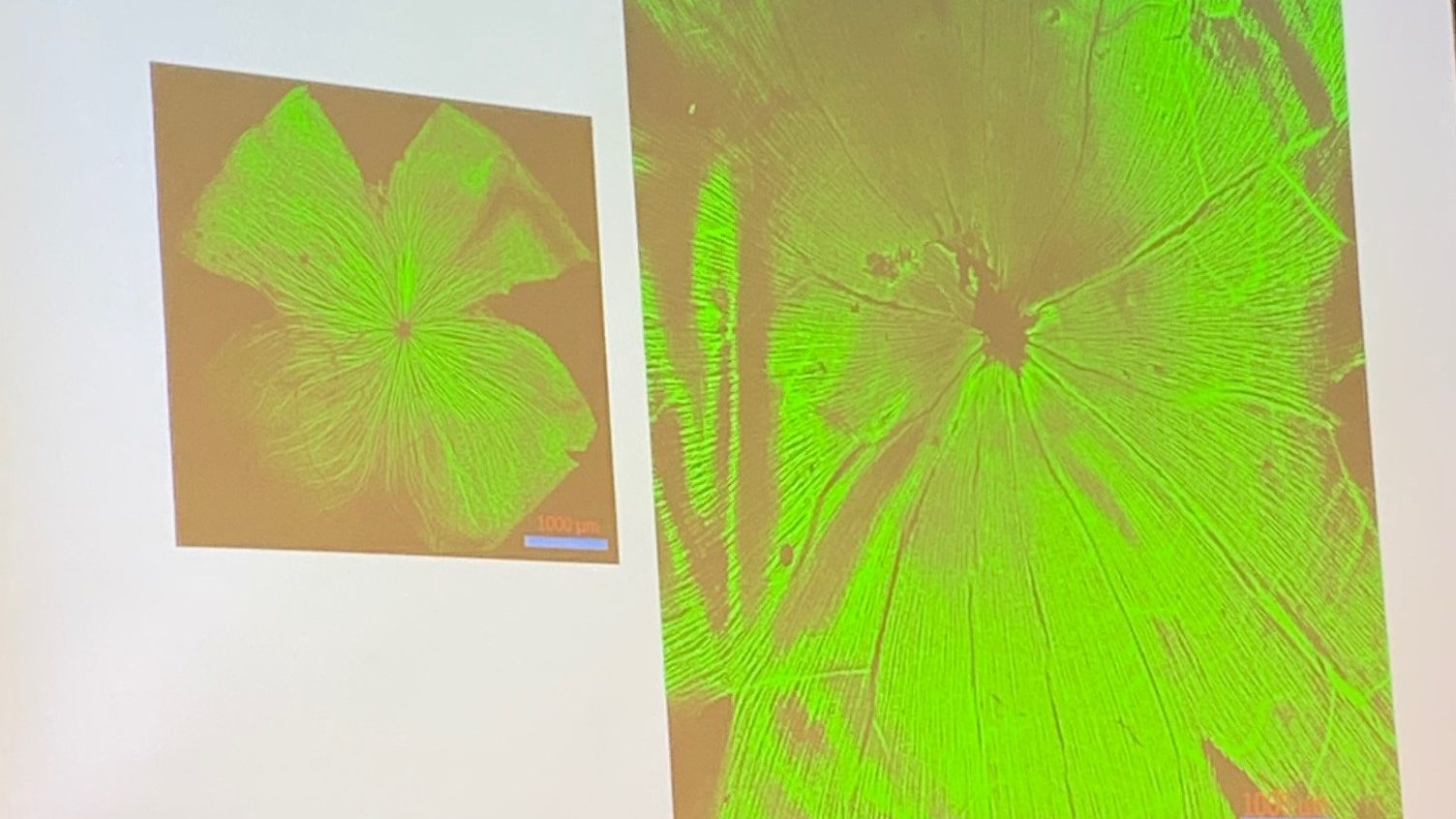
Tim Blenkinsop (Icahn School of Medicine at Mt. Sinai, USA) talked about human eye organoid models of iris development and the hope that the iris specification model his team developed will help discover the mechanisms leading to aniridia.
Brian Brooks (National Eye Institute, NIH, USA) presented about using “reprogrammed” skin cells (called “induced pluripotent stem cells” or iPSCs) to understand the relationship between the pigmented cells at the eye and foveal development.
Jennifer Elisseeff (Johns Hopkins University, USA) discussed the challenging task of trying to build a synthetic cornea and talked about new biomaterials that are promising.
Kevin Gregory-Evans (University of British Columbia, Canada) discussed the identification of a new molecule that may prevent premature cell death in tissue transplantation.
Jim Lauderdale (University of Georgia, USA) provided a video presentation focusing on a new animal model—a genetically engineered brown anole. Unlike mice and other animal models used in research, these lizards do have a fovea and therefore may be an excellent animal model for understanding foveal development in humans.
Xiaorong Liu (University of Virginia, USA) talked about a new imaging technique, called the vis-OCTF, used for better diagnosis of neural damage in eye diseases. She also highlighted work on a new animal model for research—the tree shrew, which has larger eyes than mice and good motion vision.
Awe-inspiring Complexity of the Eye
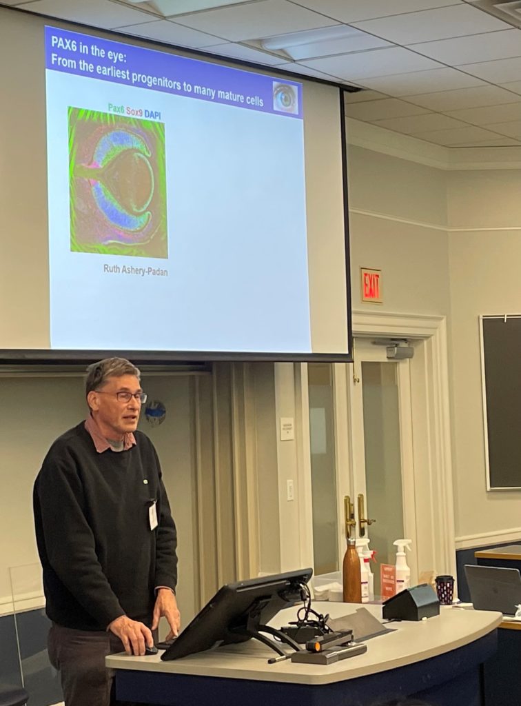
Ruth Ashery-Padan (Tel-Aviv University, Israel) presented new information regarding development of the retinal pigmented epithelium, which is a tissue essential for retinal function in the eye.
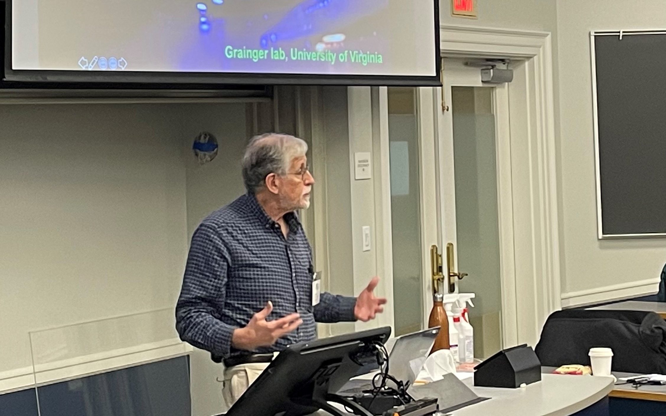
Robert Grainger (University of Virginia, USA) discussed his work using aniridic frogs to gain unprecedented insights into both normal development of the eye and the earliest stages of aniridia.
Cheryl Gregory-Evans (University of British Columbia, Canada) presented how mutant zebrafish are being used to understand the signals that support normal iris development. This is an effort to understand why defects in PAX6 result in such a wide variety of abnormalities.
Heike Kröger (University of Georgia, USA) discussed how she investigates the way cone photoreceptors develop to explore new therapies for patients who suffer from foveal hypoplasia, including cone-rod-dystrophies, achromatopsia, and aniridia.
Salil Lachke (University of Delaware, USA) described his team’s development of a new tool, called iSyTE, and how this tool was used to uncover several new proteins that are present in the eye as it forms. These data help us understand the complex mechanisms that control PAX6 production in normal eye development and uncover potential new targets for restoring optimal levels of PAX6.
A common theme for all presentations was the amazing complexity of the eye and the visual system. With the sheer number of developmental steps that must go right for vision to occur, it is remarkable that humans can see anything at all! Research into understanding the development of the eye is therefore also highly complex and technical.
Following are overviews of presentations focusing on the development and complexity of the eye:

Gratitude
As the symposium ended, patient group representatives agreed that one theme stood out: overwhelming gratitude. We are so thankful for the work of these brilliant scientists and clinicians and for the research teams with whom they work. Scientists and medical professionals agreed that this scientific symposium was a unique opportunity to present their research, discuss their activities with other professionals, and generate new ideas and collaborations. All participants agree that research provides great hope for the future.
Aniridia North America is grateful for the generous funding and support for this symposium from the University of Virginia, the Vision for Tomorrow Foundation, the University of Georgia, and the International WAGR Syndrome Association.
Author
Janelle Collins, ANA Board Member at Large
Reviewers
Robert Grainger, PhD
Jim Lauderdale, PhD
Peter Netland, PhD, MD
Kelly Trout, BSN, RN
January 2022
Wow! Overwhelmed to see so much research focusing in and around Aniridia. So many different and exciting pathways – you can’t help but think we’re on the cusp of unlocking something to get us one step closer! Thank you for sharing all of this information and a huge thank you to all the researchers and their teams!
“we’re on the cusp of unlocking something…” Yes! That’s how it definitely feels. So many different and exciting pathways indeed!